Anti-Inflammatory Diet

All health care starts with diet. My recommendations for a healthy diet are here:
Anti-Inflammatory Diet and Lifestyle.
There are over 190 articles on diet, inflammation and disease on this blog
(find topics using search [upper left] or index [lower right]), and
more articles by Prof. Ayers on Suite101 .

Showing posts with label lupus. Show all posts

Tuesday, May 27, 2014

Metformin, Antibiotic with Autoimmune Side Effects

----The other 200 posts are here----
Metformin
Major points linked in this article:
  • Metformin is commonly used in the treatment of diabetes.
  • Metformin is structurally and chemically related to arginine, guanine and Canavanine.
  • Side effects of Metformin include GI upset and autoimmune lupus (same with Canavanine.)
  • Metformin also kills bacteria, i.e. it is an antibiotic.
  • Many pharmaceuticals, e.g. statins, were first identified as antibiotics produced by fungi.
  • Antibiotics select for antibiotic resistance genes, i.e. essential bacterial genes that have mutated to no longer be inactivated by antibiotics.
  • New antibiotic resistance genes are combined with other resistance genes on multiple resistance plasmids that are transferred as a group.
  • Because of its wide use, resistance to Metformin (and statins) as an antibiotic probably already exists and has been incorporated into multiple drug resistance plasmids.
  • Many common pharmaceuticals are also antibiotics and probably select for multiple drug resistance.
  • A major contributor to multiple drug resistance, “super bugs”, and the rapid loss of efficacy of antibiotics is the over use of pharmaceuticals in general, in addition to the specific abuse of antibiotics designed to kill pathogens.
Metformin is a Good Anti-Diabetic, but...
Arginine
Metformin is the treatment of choice for type 2 diabetes and yet, like many other common drugs, the full extent of its impact on the body (and the body’s essential microbiome of bacteria and fungi) has not been studied.  This article should not be seen as a criticism of the pharmacological efficacy of Metformin in lowering blood sugar.  The point here is that Metformin alters gut flora and its major pharmacological impact may result from alteration of the gut flora and not direct action on cells of body organs.  Metformin, because of its structure and size would be expected to act relatively indiscriminately in numerous cell functions, but I don't think that these interactions are as important as the impact on gut flora.  Metformin has all of the properties of an antibiotic selected to lower blood sugar and have limited side effects.  It would not be expected to cause a dramatic increase in autoimmunity, because diabetics already have elevated autoimmunity and associated deficiencies in gut flora.

Metformin is a Diguanide
 I previously explored the interesting properties of Metformin in my laboratory and through computer modeling experiments, and found it would react with many cellular enzymes and receptors similarly to the amino acid arginine.  This was no surprise, since the working end of arginine is a guanide and Metformin is a Siamese twin of guanides, i.e. a biguanide.  I might as well also say that another guanide, Canavanine, a toxic, antimicrobial phytoalexin in bean sprouts, has similar properties.
Canavanine

Phytochemicals as Antibiotics


I have studied (and written about) the natural plant antibiotics, phytoalexins, in legumes, and particularly in soy beans, so I would expect all of the chemicals, (a.k.a. phytochemicals or “antioxidants”) extracted from plants, e.g. alkaloids, polyphenols and essential oils, to kill bacteria and be toxic to human cells.  The selective advantage to plants in producing phytochemicals is the antibiotic activity of those chemicals.  Pathogens that have adapted for growth on one species of plant have resistance genes to that plant’s phytoalexins.  Thus, bacterial genes for resistance to the antibiotic activity of drugs derived from phytochemicals are common in nature and broad use of these drugs merely selects for the transfer of these genes to gut flora.
Canavanine and Lupus
What put together more pieces of the gut flora/antibiotic/autoimmune disease puzzle for me, was coming across Dr. Loren Cordain's recent reiteration of the toxicity of legumes and his singular example of Canavanine from alfalfa sprouts as a contributor to the autoimmune disease, lupus.  When I looked up the structure of Canavanine and found it to be a guanide, I immediately started making comparisons to Metformin and was amazed to see that these chemicals share the same list of side effects focused on the gut.  Moreover, lupus is also a side effect of both Metformin and Canavanine.  It was initially surprising, that a recent study suggests that the anti-diabetic action of Metformin may result indirectly from its antibiotic effects on gut flora.  I now expect that Canavanine causes lupus by killing or altering the metabolism of particular species of bacterial gut flora involved in the normal functions of the immune system, e.g. Tregs required for immune tolerance.  It is now a common observation that many pharmaceuticals act indirectly via their impact on gut flora, i.e. many pharmaceuticals are fundamentally antibiotics, and particular antibiotics can duplicate the activity of pharmaceuticals.
Pharmaceuticals Select for Multiple Antibiotic Resistance
I have one other concern about the wide use of drugs derived from phytoalexins.  Metformin can be considered one of those drugs, and just like phytoalexins, it is a potent antibiotic.  There is no difference between purified natural plant antibiotics/ phytoalexins/ polyphenols/ antioxidants and commercially synthesized antibiotics with respect to selecting for resistance.  I would expect that resistance to Metformin, as an antibiotic, has already developed in common gut flora and consequently, that multiple drug resistance plasmids from hospital pathogens now contain Metformin resistance.  Thus, I would also expect Metformin and many other pharmaceuticals to select for multiple antibiotic resistance. [An additional example is the antibiotic activity of NSAIDs on Helicobacter pylori.  I think that prevalent use of NSAIDs in many countries is responsible for the decline in H. pylori.]


Friday, August 7, 2009

Cause of Autoimmunity and Allergies

Inflammation-Induced Presentation by Mannose Receptor

Speculation on how innocuous proteins become the targets of our immune systems, and result in allergies, asthma, celiac, arthritis, lupus, and other inflammatory autoimmune diseases.

Inflammation

It all starts with chronic inflammation. In most cases diet is the predominant source of inflammation, but infections (bacterial, viral, fungal) may also contribute. Inflammation sets the stage for faulty processing of proteins at the focal location where lymphocytes and antigen-presenting cells are congregating.

Antigen Presentation

At this point a major mistake occurs. Cells that have been alerted to danger by inflammatory cytokines, start to internalize and process proteins in the vicinity, so that peptide fragments of the “antigens” can be displayed on their surfaces in the clutches of major histocompatibility complex proteins. The problem arises when self proteins are internalized, processed and displayed as candidate peptides.

Basic Triplets of All Allergens and Autoantigens

I have looked at dozens of allergens and autoantigens, and they all have one peptide sequence in common, a triplet of basic amino acids. The impetus for this article was finding this morning that one of the autoantigens for Hashimori’s thyroiditis is thyroglobulin, the serum carrier of thyroid hormones. I checked the sequence of thyroglobulin and it has two of the rare basic triplets.

Thyroiditis Autoantigen Is Thyroglobulin

I checked the Pubmed literature to see if thyroglobulin is naturally taken up by cells, since I have been trying to figure out the receptor for basic triplets. What I found was an article on the binding of thyroiditis autoantigens to mannose receptor. The idea of the paper was that the autoantigens in this case were heavily glycosylated and maybe the mannose receptor that is involved in antigen presentation of glycosylated antigens, would bind the selected autoantigens as well. I didn’t get much past the title of the paper.

Candidates for Allergen/Autoantigen Receptor

What I put together was the fact that antigen presenting cells use mannose receptor to internalize antigens. This places this receptor in the right place and the right time to be a receptor for basic triplets. But what would a basic triple binding protein domain look like and could mannose receptor be a candidate?

Structure of Thyroglobulin

I downloaded a structure for mannose receptor and began looking for a surface region that would bind carbohydrates, aromatics and basic amino acids. The critical amino acids in all of these cases are aromatic amino acids, phenylalanine, tyrosine and tryptophan. Tryptophan is at the heart of most carbohydrate binding proteins. Since the mannose receptor is a member of the carbohydrate-binding lectins, I expected to find tryptophans on the surface of the mannose receptor. I just looked for the tryptophan that binds the carbohydrates. It was exactly as I predicted. The mannose receptor should be able to form very stable tryptophan/arginine-like ladders with basic triplets.

I made a figure of the mannose receptor bound to a carbohydrate (red and grey). One of the exposed tryptophans (yellow) is bound to the carbohydrate. Several other tryptophans could be exposed and oriented toward a basic amino acid spread over the surface of the tryptophan in place of the carbohydrate. A similar kind of structure is used by importin to transport proteins with nuclear localization sites (NLS, basic quartets) into the nucleus. Many proteins with NLSs are also autoantigens, e.g. lupus.

I think that the peculiar circumstances that lead to allergy and autoimmunity result in the binding of self-proteins or allergens to mannose receptor and result in antibody production.

Why the Mannose Receptor?

Why does the mannose receptor make mistakes? The exposed tryptophan may also bind numerous plant products. In fact, the phytochemicals, e.g. alkaloids, flavonoids and terpenoids, are an abundant and varied group of chemicals that would bind to the exposed tryptophan of the mannose receptor and compete for binding with basic triplets. Protection offered by plant “anti-oxidants” may be due in part to this activity.

Wednesday, June 24, 2009

The Cause of Allegies and Autoimmune Diseases

Keyhole Limpet Hemocyanin (KLH): Internalized Antigen

Scanning the literature for a common protein that can be used as an experimental antigen, it becomes quickly obvious that a favorite is KLH. This would seem to be an odd choice -- why a keyhole limpet protein? But that is the wrong question.

Why is KLH such a good antigen, i.e. why is it readily presented to the host immune system? If you have been reading my posts, you might be thinking about triplets of basic amino acids and that is the answer.

As soon as I remembered the prominent use of KLH as an antigen, I checked the NCBI protein database and immediately found an unusual KKK (triple lysine) near the amino terminus of hemocyanin II ( it comes in two pieces). This triplet explains why KLH is such a good experimental antigen, because it is internalized into antigen presenting cells by its strong heparin-binding domain. Other components, adjuvants, are typically added to the KLH for injection to make sure that a strong local inflammation occurs.

Autoantigens Have Strong Heparin-Binding Triplet

I also learned that Hashimoto’s thyroiditis is an autoimmune disease mediated by the autoantigen thyroid peroxidase. A quick search reveals that thyroid peroxidase is an autoantigen, because it also has a triplet of basic amino acids that can enhance presentation under inflammatory conditions. Grave’s disease of hyperthyroidism is an autoimmune disease in which the thyroid receptor (with a basic triplet) is an autoantigen. The same kind of triplet of basic amino acids was found when I searched today for fire ant antigens and mosquito antigens.

I have also looked for the triplets in protein databases. The triplets are rare in cytoplasmic and extracellular proteins. The proteins that have triplets are usually identified as autoantigens in some disease. The triplets are common in nuclear proteins, since heparin-binding and nucleic acid-binding share the same basic amino acid domains. The nuclear internalization signal also results in rapid cellular internalization, e.g. HIV-TAT, heparanase, IGF-binding proteins. Nuclear proteins are common autoantigens in lupus.

Inflammation Plus Heparin-Binding Internalization: Allergy, Autoimmunity

Chronic inflammation can produce antibodies against proteins (foreign or self) with strong heparin-binding domains (triplets or sometimes neighboring pairs of basic amino acids, lysine or arginine). The generalization explains why particular proteins in pollens, foods, insects, pets, mites, asthma, MS, lupus, celiac, etc. produce antibody responses.

Saturday, March 14, 2009

Enteroviruses, Autoimmunity, Diabetes

Insulin-producing cells of the pancreases of diabetics have been found to harbor viruses common to the gut. Antibodies to the virus coat proteins also bind to pancreas proteins.

A recent paper, referenced below, shows that in a collection of samples from the pancreases of individuals that had been diagnosed with type I diabetes less than one year prior to the sampling, insulin producing cells are also infected with enterovirus. The same association between enterovirus infection was found to a lesser extent in type II diabetics, but not in non-diabetic controls.

Enteroviruses have been repeated associated with diabetes over the last decade and antigenic determinants of the enterovirus protein coat also bind, i.e. cross react, with antigenic determinants of human cellular proteins.

I examined the enterovirus coat protein, VP1, and found the same three amino acid sequence (three basic amino acids, lysine [K] or arginine [R], highlighted) that I also found in all allergens (peanut, ragweed, dust mite, bee venom) and autoantigens of autoimmune diseases (lupus, MS), and is associated with heparan sulfate-based internalization and presentation of protein immunogens. This observation is consistent with my hypothesis that inflammation plus the presence of one of these proteins, results in production of B and T lymphocytes specific for antigenic determinants on the surface of the immunogen protein. Note that the antigenic determinants usually do not include the three basic amino acid sequence, e.g. RRK, that is involved in uptake and presentation of the protein.

VP1 [Human enterovirus B]
HVINYHTRSESSVENFMGRAACVYIAQYATEKVNDELDR
YTNWEITTRQVAQLRRKLEMFTYMRFDLEVTFVITSSQR
TSTTYASDSPPLTHQVM

reference:
Richardson SJ, Willcox A, Bone AJ, Foulis AK, Morgan NG. 2009. The prevalence of enteroviral capsid protein vp1 immunostaining in pancreatic islets in human type 1 diabetes. Diabetologia. Mar 6. [Epub ahead of print]

Wednesday, November 26, 2008

CIDP, Inflammation and Autoantigens

Chronic Inflammatory Demyelinating Polyneuropathy (CIDP) is chronic, relapsing autoimmune disease in which the immune system attacks the insulating myelin sheaths of nerves in the limbs. The result is numbness and pain. An acute version of this myelin compromising disease is Guillain-Barre syndrome (GBS) typically initiated by Campylobacter jejuni infection. A related disease of brain tissue is multiple sclerosis (MS). In each of these cases antibodies bind to protein or glycolipid antigens unique to myelin and target the attack by lymphocytes.

Autoimmune diseases represent a disruption of the normal function of the immune system that arises from a breakdown of the self/non-self tolerance process. Immune responses by antibodies or lymphocytes is normally reserved for molecules that don’t exist in the body, i.e. self. Differentiation of lymphocytes in the thymus or bone marrow involves systematic elimination of all lymphocytes that interact with self antigens. There are, however, self antigens that are not normally encountered by the immune system, e.g. nuclear antigens, brain antigens, since these antigens are only excluded from the cytoplasm or result from specialized differentiation. Thus, you can find in mystery novels the use of letters coated with powdered brain antigen to slowly kill targets by induction of a lethal autoimmune disease.

The self antigens that are the typical targets of the CIDP, GB and MS are gangliosides, myelin glycolipid also produced by C. jejuni, and basic proteins such as peripheral myelin protein 22. The glycolipid makes sense, because the initial exposure is during a bacterial infection that also produces inflammation. The basic proteins are a different case.

I have examined numerous self or autoantigens that are the targets for antibodies produced in autoimmune diseases and allergies. In each case the same amino acid sequence or domain is present, three basic (positively charged, arginine or lysine) amino acids. This is true for the myelin autoantigens; allergens from ragweed, mites, peanut, etc.; diabetes, lupus and arthritis.

Here is the example of the CIDP autoantigen, peripheral myelin protein 22:
MLLLLLSIIVLHVAVLVLLFVSTIVSQW
IVGNGHATDLWQNCSTSSSGNVHHCF
SSSPNEWLQSVQATMILSIIFSILSLFLF
FCQLFTLTKGGRFYITGIFQILAGLCVM
SAAAIYTVRHPEWHLNSDYSYGFAYIL
AWVAFPLALLSGVIYVILRKRE

The three basic amino acids, RKR, are at the end of the protein amino acid sequence.

A dramatic instance of tissue damage caused by physical injury or infection, is superimposed on chronic dietary inflammation to provide the environment for an inappropriate response of the immune system to a self or identical foreign antigen. The only proteins that seem to be presented in this way to the immune system are those with the strong heparin-binding domain, BBB (B = basic amino acid). This indicates that perhaps the reduction in heparin production that accompanies inflammation, may be responsible for the aberrant immune response. It is also noteworthy that heparin and anti-inflammatory agents can reduce the symptoms of many autoimmune diseases.

There are many complex therapies to deal with the symptoms of these autoimmune diseases. In all of these cases, one of the most effective approaches is an anti-inflammatory diet and lifestyle. A novel additional approach is helminth therapy, infection with parasites to suppress the T-lymphocyte autoimmune attack. This approach is effective with MS, and I think would be an excellent, relatively safe approach to be examined for the other myelin autoimmune diseases.

Tuesday, November 11, 2008

Bee Sting Allergy

Typical. I started to write an article on leukotrienes, the inflammatory derivatives of the omega-6 fatty acid, arachidonic acid, but ran across another powerful example to test my hypothesis to explain the cause of allergies. The leukotriene article will have to wait till another day.

Wikipedia is my source of choice for up-to-date summaries of biomedical information. I queried “leukotrienes” and immediately ran across the original name for these inflammatory compounds, “slow reacting substance of anaphylaxis”. I was initially distracted by the classic experimental use of snake venom and histamine to induce leukotriene production. Snake venom has the same enzyme, PLA2, as brown recluse spider venom (subject of a previous article) and honey bee venom, that releases arachidonic acid (ADA). ADA is an omega-6 fatty acid that is the starting material for inflammatory prostaglandins and leukotrienes.

The mention of honey bee venom in the Wikipedia article on anaphylaxis sent me on a quick check of the structure and sequence of the honey bee allergen. I initially found that the major allergen is a hyaluronidase. I quickly searched for a three amino acid sequence that I predicted would make it an allergen. It was just where I expected to find it. About two thirds of the way along the amino acid sequence I found, -TTSRKKVLP-. Three basic amino acids together, in this case -RKK-, argininine-lysine-lysine, form a strong heparin-binding domain, that I believe takes proteins into cells and during inflammation primes the immune system for allergic responses.

I have found the same strong heparin-binding domain associated with allergens of ragweed, dust mites and peanuts. The principal autoantigens of autoimmune diseases, such as lupus, celiac, etc. also display the same unusual sequences. In lupus, or example, nuclear proteins with the internalization signal provided by nucleic acid-binding domains (and nuclear localization signas) are autoantigens. This pattern is found with all allergens that I have examined. There are a few apparent exceptions, but in all of these cases, there is a closely related allergen from a related source that has the expected strong heparin-binding domain. It appears that in these cases, the less common allergen provides the initial exposure during the presentation phase of high inflammation, and the allergy is maintained by subsequent exposure to the more common allergen. After the establishment of the allergy, the strong heparin-binding domain is no longer needed, because antibodies bind to other parts of homologous allergenic proteins for internalization.

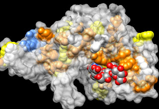
Just for fun, I have illustrated the honey bee allergen, hyaluronidase, to show both its strong heparin-binding domain (blue) along with its substrate hyaluronan (grey and red). Note that the substrate sugars are in the slot of the active site, which is lined with orange and yellow aromatic amino acids that provide flat, hydrophobic binding platforms for each sugar.

After this little distraction to provide further support for my explanation of the cause of allergies, I have to get back to looking at the role of leukotrienes in anaphylaxis, COPD, asthma and other inflammatory diseases.